Radiographic Sins of Idiopathic Intracranial Hypertension
Symptoms of Idiopathic Intracranial Hypertension (IIH) include:
Headaches (IIH)
There are three hallmark symptoms associated with this disease, with headaches being the most common. While there may not be a specific headache pattern, patients often describe pain behind the eyes or in the back of the head, that worsens when lying flat or first waking up. Migraine type headaches can also be present, and many patients may in fact be treated for migraine headaches
Changes in Vision (IIH)
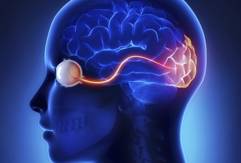
The second most common symptom is visual changes. Experiencing blurry vision or vision loss when the head is bent down is particularly associated with intracranial hypertension. Other symptoms have been described such as seeing flashes of light or having double vision.
An eye doctor can look at the back of the eyes, the optic discs, to see if there is swelling of the nerve. When swelling occurs, this is called papilledema. They will also perform visual field testing to check for an enlarged blind spot. Even if no signs are present, one can still have the disease.
Pulsatile Tinnitus (IIH)
The symptom that is most suggestive of raised intracranial pressure is pulsatile tinnitus. Pulsatile tinnitus is a pulsating sound that comes and goes with the heartbeat and is worst at night. Patients experiencing pulsatile tinnitus complain of hearing their own blood flow, often described as a whooshing sound.
Pulsatile tinnitus can occur in one or both ears. If this symptom is present, one should think about IIH.
Nausea and dizziness can also be reported. Some people have a diagnosis of high blood pressure as well, or are on medications for high blood pressure.
IIH Diagnosis
Many people struggle to get the correct diagnosis of idiopathic intracranial hypertension. The diagnosis is characterized by elevated intracranial pressures. It is a disease that is likely underdiagnosed and affects predominantly women of childbearing age. There are various tests including imaging, that can help diagnose IIH
Radiographic Signs (IIH)
Elevated pressures can be detected on a MR brain image or a more specialized MR Venogram. Here are some of the radiographic signs listed from most common to least common.
The most common sign is bilateral transverse sigmoid sinus stenosis (TSSS). It is seen in over 90% of patients with IIH, but it is really rare when the disease is not present.
Bilateral transverse sigmoid sinus stenosis is the most common sign radiologically sign for IIH. In one study bilateral TSSS was seen in over 90% of MR Venograms of IIH patients compared to 3% of controls.
That means It is rare to have TSSS if you don’t have the disease. What’s not rare, is that transverse sigmoid sinus stenosis is not reported. Many patients may not receive a MR Venogram in the first place.
Fortunately, a lot of patients will receive MRI brain imaging to look for brain tumors. TSSS can be detected on contrast enhanced MR brain sequences as well, but someone with expertise needs to look at the images.
The Tri-State's leaders
in Cerebrovascular treatments.
“Empty sella” or “partially empty sella” is seen in 70-80% of patients, where the pituitary gland gets compressed by increased pressures in the brain to the edges of the bony space called the sella. It’s termed a secondary empty sella syndrome. When empty sella syndrome occurs, people usually don’t experience any abnormal pituitary function as best as we know.
Some patients may receive a MRI of the eyes, called an MRI orbit. Flattening of the eye balls can be seen here and twisting of the optic nerve, along with other findings. If these conditions are present, it is likely that the patient has already seen an eye doctor for issues with their vision.
Other findings to look for and which have been found to have an association with IIH, are quite technical and maybe reported as “aberrant arachnoid granulations”, “brain herniation into arachnoid granulations” Slit like ventricles in itself are a less reliable sign of IIH.
And finally, patients with Chiari malformation type 1 should be evaluated for IIH, particularly if they also hear whooshing sounds.
The diagnosis remains a clinical diagnosis with some patients experiencing all symptoms and signs. Others may show few symptoms or symptoms that are not known to be associated with the disease, such as brain fog.
It is possible to have imaging findings and not experience symptoms.
Lumbar Puncture (IIH)
It is necessary to determine if the pressures in the spinal fluid are elevated. This is usually done by performing a lumbar puncture also known as a spinal tap. During the procedure a small needle is put into the cerebrospinal fluid and the pressure is measured.
The procedure is similar to an ”epidural during childbirth” except when testing for IIH no medication is injected, and there won’t be any numbness or weakness.
Treatment Options (IIH)
There are many different physician specialties that have touch points with these patients. Treatment usually involves medication and weight loss. For patients who do not experience relief with those measures, there are other treatment options.
For example in those with pulsatile tinnitus and a transverse sigmoid sinus stenosis, a venous stent can provide relief from venous congestion. This reduces pressure leading to improvement of symptoms.
This disease is still being researched and there is much more that we need to understand about it. For the time being, it is incurable but many treatments are available to help control the symptoms.

About Dr. Dorothea Altschul
Dr. Dorothea Altschul is an accomplished neurointerventionalist in North Jersey and is the Clinical Director of Endovascular Services at Neurosurgeons of New Jersey, practicing out of their Ridgewood office located on East Ridgewood Avenue.


